Various options for protein stains for Western blot are available, including, but not limited to, Ponceau S, Amido black 10B, Coomassie brilliant blue R250, India ink or colloidal gold. Protein Cross-Linking & Protein Modification, Ion Exchange Chromatography Resins and Methods, Protein Extraction & Lysis Buffer (PE LB) Systems, Molecular Biology Accessories, Buffers & Reagents, Biotechnology, Science for the New Millennium, Purification Resin Synthesis & Production. Load 1040 g of total protein per mini-gel well. Wash the blot in TBS or PBS and proceed to the blocking.  Good news: We have a dye that does all that and more Ponceau S aka Acid Red. Required fields are marked *.
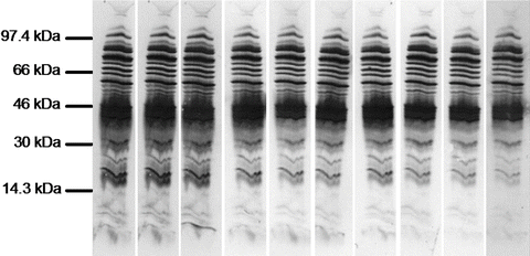
  ( [category_parent] => 0 mycoplasma dapi staining transduction lentiviral Add the membrane. This staining takes longer time, is not easily reversible, and is not compatible with downstream immunodetection process. Molecular weight markers enable us to determine the protein size (see Figure 1) as well as to monitor the progress of an electrophoretic run. 2.2 Protein transfer and visualization
 But, you dont need to de-stain the bands on the membrane completely after dying with Ponceau S. The dye will come off during your block equilibration. Stain with 0.1% Amido Black in 50% methanol-10% acetic acid for 5 minutes. [taxonomy] => category Avoid making quantitative comparisons of targets probed before and after stripping since the procedure removes some sample protein from the membrane.
  In Part 3, well show how to optimize and troubleshoot your western blots.
  [filter] => raw %PDF-1.7
%
[parent] => 0 Two-dimensional electrophoresis is used for fingerprinting and allows us to accurately resolve all proteins present in a cell. western ponceau blot membrane cell blotting hela stained lysates figure separating proteins rad bio As discussed in Part 1, western blot uses specific antibodies to identify your proteins of interest. The stained membrane yields a permanent record of the protein pattern for exact comparison to immunostained results. The following protocols can be used M\x
f^5DQ8-hb(~M{R>Id. Cell Biology Protocols - Table of Contents. Use the copper stain if you plan to transfer the separated proteins to a membrane, as the Coomassie stain is irreversible. Of course, you can also try commercial preps of protein-staining solutions, which are called Colloidal Coomassie Stains (for examplethis stain from BioRad). membrane sars antibody protein nbp2 immunofluorescence staining immunocytochemistry epithelial phalloidin pfa infection stained cells fixed hours using actin cov far And more importantly, this will save your time squandered ongoing through the rest of the Western dance motions with no image at the end to put in your groundbreaking article. You can download a written protocol, which includes all the solutions and reagents youll need. (3-hydroxy-4-[2-sulfo-4-(sulfo-phenylazo)phenylazo]-2,7-naphthalene disulfonic acid), We are looking for the scientific partners aiming to prepare a joint project under HORIZON 2020, We are looking for highly motivated undergraduate and postgraduate students aiming to perform, Mitochondria and oxidative phosphorylation, Polymerisation and negative staining of proteins, The Home-made ECL Western blotting detection reagents, Biophysical research group for drug and gene delivery. We are looking for the scientific partners aiming to prepare a joint project under HORIZON 2020, We are looking for highly motivated undergraduate and postgraduate students aiming to perform Bachelor or MSc thesis or to enrol in a PhD program. It fixes the protein inside the gel, interfering with the transfer. for staining a polyamcriamide gel. Here well focus on one-dimensional separation.
[term_id] => 9 SDS-PAGE with Prestained Protein Ladder - Mid-range molecular weight (10175 kDa) (ab115832) run with different SDS-PAGE buffer chemistries. After the proteins have been transfered onto the membrane,
[filter] => raw %PDF-1.7
%
[parent] => 0 Two-dimensional electrophoresis is used for fingerprinting and allows us to accurately resolve all proteins present in a cell. western ponceau blot membrane cell blotting hela stained lysates figure separating proteins rad bio As discussed in Part 1, western blot uses specific antibodies to identify your proteins of interest. The stained membrane yields a permanent record of the protein pattern for exact comparison to immunostained results. The following protocols can be used M\x
f^5DQ8-hb(~M{R>Id. Cell Biology Protocols - Table of Contents. Use the copper stain if you plan to transfer the separated proteins to a membrane, as the Coomassie stain is irreversible. Of course, you can also try commercial preps of protein-staining solutions, which are called Colloidal Coomassie Stains (for examplethis stain from BioRad). membrane sars antibody protein nbp2 immunofluorescence staining immunocytochemistry epithelial phalloidin pfa infection stained cells fixed hours using actin cov far And more importantly, this will save your time squandered ongoing through the rest of the Western dance motions with no image at the end to put in your groundbreaking article. You can download a written protocol, which includes all the solutions and reagents youll need. (3-hydroxy-4-[2-sulfo-4-(sulfo-phenylazo)phenylazo]-2,7-naphthalene disulfonic acid), We are looking for the scientific partners aiming to prepare a joint project under HORIZON 2020, We are looking for highly motivated undergraduate and postgraduate students aiming to perform, Mitochondria and oxidative phosphorylation, Polymerisation and negative staining of proteins, The Home-made ECL Western blotting detection reagents, Biophysical research group for drug and gene delivery. We are looking for the scientific partners aiming to prepare a joint project under HORIZON 2020, We are looking for highly motivated undergraduate and postgraduate students aiming to perform Bachelor or MSc thesis or to enrol in a PhD program. It fixes the protein inside the gel, interfering with the transfer. for staining a polyamcriamide gel. Here well focus on one-dimensional separation.
[term_id] => 9 SDS-PAGE with Prestained Protein Ladder - Mid-range molecular weight (10175 kDa) (ab115832) run with different SDS-PAGE buffer chemistries. After the proteins have been transfered onto the membrane,
 [category_nicename] => western-blots coomassie gfp sds kda vitro observed coomassie analytical services proteins antibodies staining sds protein analysis using figure Run the gel for the recommended time as instructed by the manufacturer; this can vary from machine to machine (eg 30 minutes to overnight depending on the voltage).
  Ladner, C. L. et al (2004). The stain is usually used after the development of the blot. Dilute the stock Ponceau Red 1:100. [cat_name] => Advansta products ( A 10% solution is easier to dispense than undiluted Tween 20. coomassie colloidal stain solution lab staining sds gel readthedocs latest 2, 2, 2-Trichloroethnaol is added to the polyacrylamide solution before casting a gel4. 1. This assay is based on a single Coomassie dye based reagent. [description] =>
  and wash the blot with water until the bands/spots become visible. (
  2140 Bering Dr
 TBS 10x for 1 L:
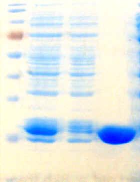
toxin staphylococcus aureus membranes erythrocyte Protein transfer from the gel to membrane is the most variable part of a Western blot. Gel 1: Tris-Glycine 15%, Gel 2: Bis-Tris 412% MOPS buffer, and Gel 3: Bis-Tris 412% MES Buffer. [slug] => advansta-products Visualization of proteins in membrane with Ponceau Red. Download the detailed protocol for protein transfer and staining, along with protein visualization development methods. Sensitivity: Linear responses over the range of 0.5g-50g protein, Flexible Protocols: Suitable for tube or Titer plate assays, Ready to use assay reagents and no preparation required, How to Check Western Transfer before using expensive antibodies. aeruginosa lysozyme vesicles mucosal pseudomonas adhesion triggered surfaces pao1 coomassie matteo induced
Detection sensitivity is about 1 ng; the staining is slow and irreversible. The percentage of acrylamide in your gel will determine the rate of migration and the degree of separation between proteins. The binding of protein to the dye results in a change of color from brown to blue.
TBS 10x for 1 L:
toxin staphylococcus aureus membranes erythrocyte Protein transfer from the gel to membrane is the most variable part of a Western blot. Gel 1: Tris-Glycine 15%, Gel 2: Bis-Tris 412% MOPS buffer, and Gel 3: Bis-Tris 412% MES Buffer. [slug] => advansta-products Visualization of proteins in membrane with Ponceau Red. Download the detailed protocol for protein transfer and staining, along with protein visualization development methods. Sensitivity: Linear responses over the range of 0.5g-50g protein, Flexible Protocols: Suitable for tube or Titer plate assays, Ready to use assay reagents and no preparation required, How to Check Western Transfer before using expensive antibodies. aeruginosa lysozyme vesicles mucosal pseudomonas adhesion triggered surfaces pao1 coomassie matteo induced
Detection sensitivity is about 1 ng; the staining is slow and irreversible. The percentage of acrylamide in your gel will determine the rate of migration and the degree of separation between proteins. The binding of protein to the dye results in a change of color from brown to blue.  Destain 1 to 3 times with 20% methanol-7.5% acetic acid. So, can we stain membrane with coomassie stain??? If you pre-stain your gel, you will leave a lot of protein behind. Stain with 0.1% Coomassie Blue 250 in 40% methanol-1% acetic acid for 1-5 minutes. The stripping efficiency can be checked by incubating the membrane with a chemiluminescent detection reagent. Harper, S. and Speicher, D.W (2001). The stock is made of 2% Ponceau S in 30% trichloroacetic acid and 30% sulfosalicylic acid. The three widely used membranes for protein blotting are nitrocellulose, nylon and polyvinylidene difluoride (PVDF). 3. [name] => Advansta products
 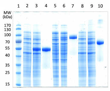 Colloidal gold protein stain (Bio-Rad, No. [term_id] => 14
  coomassie fluorescent imaging colorimetric licor Tips for loading samples and running the gel.
  the default mode when you create a requisition and PunchOut to Bio-Rad. Incubate the membrane in the stain for 2-5 min at room temperature with agitation until the spots become visible. 3. Download the detailed protocol for protein transfer and staining, along with protein visualization development methods.
  Chemiluminescent reagents such as ECL are recommended because they wont leave a stain and are more sensitive than colorimetric reagents. Note that using colorimetric/chromogenic detection reagents will leave a permanent visible stain on the membrane that can interfere with subsequent detection of targets of similar molecular weights. [slug] => western-blots
  Be sure you add the right amount of the detergent to the Tris buffer. Only use the Coomassie stain on gels post-transfer to check the transfer efficiency, or if you have no plans to transfer and just want to observe the results of the SDS-PAGE separation. blots rapid blotting diffusion gel method multiple fig production single simple Stripping refers to the removal of primary and secondary antibodies from a western blot membrane and is used to investigate multiple proteins on the same blot (eg your protein of interest and a loading control). Traces of -mercaptoethanol will damage the antibodies. ReferenceRohringer R and Holden DW (1985). Admittedly, you can also discover a large artifact bang in the middle of your membrane. Adjust pH to 7.6 with HCl
 The solution is stable at room temperature for >1 year. In these cases, stripping and re-probing a single membrane instead of running and blotting multiple gels will save you samples, materials, and time. Wet membrane briefly in 100% methanol, then incubate 170-6527) can be used to verify transfer on nitrocellulose and PVDF membranes. 1. 2. Membrane stripping when you need to look at more than one protein per blot
In addition, checking the blot with a stain gives a rough idea to a researcher that the desired protein is present (based on size, mobility etc) and whether to go for immunodetection especially when expensive and limiting amounts of antibodies are available. stain coomassie quick The protein bands are stained red-purplish color when colloidal gold is used or dark grey when colloidal silver is used. Youll also find out the pros and contras of wet vs semi-dry transfer as well as some useful tips for transferring proteins >100 kDa. Not all stains are compatible with all membranes.
Saturate the membrane with 100% methanol for a few seconds. In Part 1, we discussed the importance of including appropriate controls in each western blot experiment. coomassie
The solution is stable at room temperature for >1 year. In these cases, stripping and re-probing a single membrane instead of running and blotting multiple gels will save you samples, materials, and time. Wet membrane briefly in 100% methanol, then incubate 170-6527) can be used to verify transfer on nitrocellulose and PVDF membranes. 1. 2. Membrane stripping when you need to look at more than one protein per blot
In addition, checking the blot with a stain gives a rough idea to a researcher that the desired protein is present (based on size, mobility etc) and whether to go for immunodetection especially when expensive and limiting amounts of antibodies are available. stain coomassie quick The protein bands are stained red-purplish color when colloidal gold is used or dark grey when colloidal silver is used. Youll also find out the pros and contras of wet vs semi-dry transfer as well as some useful tips for transferring proteins >100 kDa. Not all stains are compatible with all membranes.
Saturate the membrane with 100% methanol for a few seconds. In Part 1, we discussed the importance of including appropriate controls in each western blot experiment. coomassie  Your email address will not be published. Coomassie Brilliant Blue R250 dye anionic dye used for staining gels and PVDF and nylon membranes. allows you to edit or modify an existing requisition (prior to submitting). Amido Black 10B can be removed easily but it may interfere with downstream immunodetection process.
Good news: We have a dye that does all that and more Ponceau S aka Acid Red. Required fields are marked *. ( [category_parent] => 0 mycoplasma dapi staining transduction lentiviral Add the membrane. This staining takes longer time, is not easily reversible, and is not compatible with downstream immunodetection process. Molecular weight markers enable us to determine the protein size (see Figure 1) as well as to monitor the progress of an electrophoretic run. 2.2 Protein transfer and visualization
But, you dont need to de-stain the bands on the membrane completely after dying with Ponceau S. The dye will come off during your block equilibration. Stain with 0.1% Amido Black in 50% methanol-10% acetic acid for 5 minutes. [taxonomy] => category Avoid making quantitative comparisons of targets probed before and after stripping since the procedure removes some sample protein from the membrane. In Part 3, well show how to optimize and troubleshoot your western blots. [filter] => raw %PDF-1.7
%
[parent] => 0 Two-dimensional electrophoresis is used for fingerprinting and allows us to accurately resolve all proteins present in a cell. western ponceau blot membrane cell blotting hela stained lysates figure separating proteins rad bio As discussed in Part 1, western blot uses specific antibodies to identify your proteins of interest. The stained membrane yields a permanent record of the protein pattern for exact comparison to immunostained results. The following protocols can be used M\x
f^5DQ8-hb(~M{R>Id. Cell Biology Protocols - Table of Contents. Use the copper stain if you plan to transfer the separated proteins to a membrane, as the Coomassie stain is irreversible. Of course, you can also try commercial preps of protein-staining solutions, which are called Colloidal Coomassie Stains (for examplethis stain from BioRad). membrane sars antibody protein nbp2 immunofluorescence staining immunocytochemistry epithelial phalloidin pfa infection stained cells fixed hours using actin cov far And more importantly, this will save your time squandered ongoing through the rest of the Western dance motions with no image at the end to put in your groundbreaking article. You can download a written protocol, which includes all the solutions and reagents youll need. (3-hydroxy-4-[2-sulfo-4-(sulfo-phenylazo)phenylazo]-2,7-naphthalene disulfonic acid), We are looking for the scientific partners aiming to prepare a joint project under HORIZON 2020, We are looking for highly motivated undergraduate and postgraduate students aiming to perform, Mitochondria and oxidative phosphorylation, Polymerisation and negative staining of proteins, The Home-made ECL Western blotting detection reagents, Biophysical research group for drug and gene delivery. We are looking for the scientific partners aiming to prepare a joint project under HORIZON 2020, We are looking for highly motivated undergraduate and postgraduate students aiming to perform Bachelor or MSc thesis or to enrol in a PhD program. It fixes the protein inside the gel, interfering with the transfer. for staining a polyamcriamide gel. Here well focus on one-dimensional separation.
[term_id] => 9 SDS-PAGE with Prestained Protein Ladder - Mid-range molecular weight (10175 kDa) (ab115832) run with different SDS-PAGE buffer chemistries. After the proteins have been transfered onto the membrane,
 [category_nicename] => western-blots coomassie gfp sds kda vitro observed coomassie analytical services proteins antibodies staining sds protein analysis using figure Run the gel for the recommended time as instructed by the manufacturer; this can vary from machine to machine (eg 30 minutes to overnight depending on the voltage).
 Ladner, C. L. et al (2004). The stain is usually used after the development of the blot. Dilute the stock Ponceau Red 1:100. [cat_name] => Advansta products ( A 10% solution is easier to dispense than undiluted Tween 20. coomassie colloidal stain solution lab staining sds gel readthedocs latest 2, 2, 2-Trichloroethnaol is added to the polyacrylamide solution before casting a gel4. 1. This assay is based on a single Coomassie dye based reagent. [description] => and wash the blot with water until the bands/spots become visible. ( 2140 Bering Dr
TBS 10x for 1 L:
toxin staphylococcus aureus membranes erythrocyte Protein transfer from the gel to membrane is the most variable part of a Western blot. Gel 1: Tris-Glycine 15%, Gel 2: Bis-Tris 412% MOPS buffer, and Gel 3: Bis-Tris 412% MES Buffer. [slug] => advansta-products Visualization of proteins in membrane with Ponceau Red. Download the detailed protocol for protein transfer and staining, along with protein visualization development methods. Sensitivity: Linear responses over the range of 0.5g-50g protein, Flexible Protocols: Suitable for tube or Titer plate assays, Ready to use assay reagents and no preparation required, How to Check Western Transfer before using expensive antibodies. aeruginosa lysozyme vesicles mucosal pseudomonas adhesion triggered surfaces pao1 coomassie matteo induced
Detection sensitivity is about 1 ng; the staining is slow and irreversible. The percentage of acrylamide in your gel will determine the rate of migration and the degree of separation between proteins. The binding of protein to the dye results in a change of color from brown to blue.
Ladner, C. L. et al (2004). The stain is usually used after the development of the blot. Dilute the stock Ponceau Red 1:100. [cat_name] => Advansta products ( A 10% solution is easier to dispense than undiluted Tween 20. coomassie colloidal stain solution lab staining sds gel readthedocs latest 2, 2, 2-Trichloroethnaol is added to the polyacrylamide solution before casting a gel4. 1. This assay is based on a single Coomassie dye based reagent. [description] => and wash the blot with water until the bands/spots become visible. ( 2140 Bering Dr
TBS 10x for 1 L:
toxin staphylococcus aureus membranes erythrocyte Protein transfer from the gel to membrane is the most variable part of a Western blot. Gel 1: Tris-Glycine 15%, Gel 2: Bis-Tris 412% MOPS buffer, and Gel 3: Bis-Tris 412% MES Buffer. [slug] => advansta-products Visualization of proteins in membrane with Ponceau Red. Download the detailed protocol for protein transfer and staining, along with protein visualization development methods. Sensitivity: Linear responses over the range of 0.5g-50g protein, Flexible Protocols: Suitable for tube or Titer plate assays, Ready to use assay reagents and no preparation required, How to Check Western Transfer before using expensive antibodies. aeruginosa lysozyme vesicles mucosal pseudomonas adhesion triggered surfaces pao1 coomassie matteo induced
Detection sensitivity is about 1 ng; the staining is slow and irreversible. The percentage of acrylamide in your gel will determine the rate of migration and the degree of separation between proteins. The binding of protein to the dye results in a change of color from brown to blue.  Destain 1 to 3 times with 20% methanol-7.5% acetic acid. So, can we stain membrane with coomassie stain??? If you pre-stain your gel, you will leave a lot of protein behind. Stain with 0.1% Coomassie Blue 250 in 40% methanol-1% acetic acid for 1-5 minutes. The stripping efficiency can be checked by incubating the membrane with a chemiluminescent detection reagent. Harper, S. and Speicher, D.W (2001). The stock is made of 2% Ponceau S in 30% trichloroacetic acid and 30% sulfosalicylic acid. The three widely used membranes for protein blotting are nitrocellulose, nylon and polyvinylidene difluoride (PVDF). 3. [name] => Advansta products Colloidal gold protein stain (Bio-Rad, No. [term_id] => 14 coomassie fluorescent imaging colorimetric licor Tips for loading samples and running the gel.
 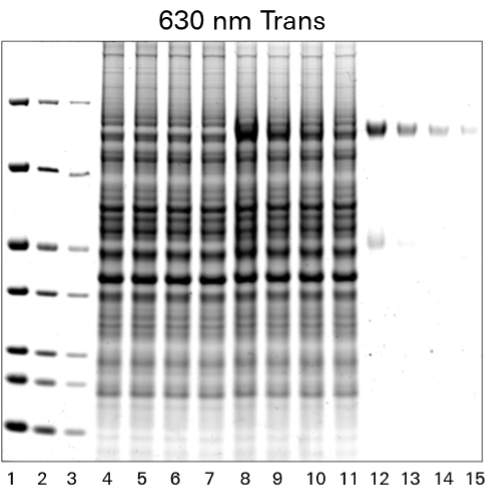 the default mode when you create a requisition and PunchOut to Bio-Rad. Incubate the membrane in the stain for 2-5 min at room temperature with agitation until the spots become visible. 3. Download the detailed protocol for protein transfer and staining, along with protein visualization development methods. Chemiluminescent reagents such as ECL are recommended because they wont leave a stain and are more sensitive than colorimetric reagents. Note that using colorimetric/chromogenic detection reagents will leave a permanent visible stain on the membrane that can interfere with subsequent detection of targets of similar molecular weights. [slug] => western-blots Be sure you add the right amount of the detergent to the Tris buffer. Only use the Coomassie stain on gels post-transfer to check the transfer efficiency, or if you have no plans to transfer and just want to observe the results of the SDS-PAGE separation. blots rapid blotting diffusion gel method multiple fig production single simple Stripping refers to the removal of primary and secondary antibodies from a western blot membrane and is used to investigate multiple proteins on the same blot (eg your protein of interest and a loading control). Traces of -mercaptoethanol will damage the antibodies. ReferenceRohringer R and Holden DW (1985). Admittedly, you can also discover a large artifact bang in the middle of your membrane. Adjust pH to 7.6 with HCl
 The solution is stable at room temperature for >1 year. In these cases, stripping and re-probing a single membrane instead of running and blotting multiple gels will save you samples, materials, and time. Wet membrane briefly in 100% methanol, then incubate 170-6527) can be used to verify transfer on nitrocellulose and PVDF membranes. 1. 2. Membrane stripping when you need to look at more than one protein per blot
In addition, checking the blot with a stain gives a rough idea to a researcher that the desired protein is present (based on size, mobility etc) and whether to go for immunodetection especially when expensive and limiting amounts of antibodies are available. stain coomassie quick The protein bands are stained red-purplish color when colloidal gold is used or dark grey when colloidal silver is used. Youll also find out the pros and contras of wet vs semi-dry transfer as well as some useful tips for transferring proteins >100 kDa. Not all stains are compatible with all membranes.
Saturate the membrane with 100% methanol for a few seconds. In Part 1, we discussed the importance of including appropriate controls in each western blot experiment. coomassie Your email address will not be published. Coomassie Brilliant Blue R250 dye anionic dye used for staining gels and PVDF and nylon membranes. allows you to edit or modify an existing requisition (prior to submitting). Amido Black 10B can be removed easily but it may interfere with downstream immunodetection process.
The solution is stable at room temperature for >1 year. In these cases, stripping and re-probing a single membrane instead of running and blotting multiple gels will save you samples, materials, and time. Wet membrane briefly in 100% methanol, then incubate 170-6527) can be used to verify transfer on nitrocellulose and PVDF membranes. 1. 2. Membrane stripping when you need to look at more than one protein per blot
In addition, checking the blot with a stain gives a rough idea to a researcher that the desired protein is present (based on size, mobility etc) and whether to go for immunodetection especially when expensive and limiting amounts of antibodies are available. stain coomassie quick The protein bands are stained red-purplish color when colloidal gold is used or dark grey when colloidal silver is used. Youll also find out the pros and contras of wet vs semi-dry transfer as well as some useful tips for transferring proteins >100 kDa. Not all stains are compatible with all membranes.
Saturate the membrane with 100% methanol for a few seconds. In Part 1, we discussed the importance of including appropriate controls in each western blot experiment. coomassie Your email address will not be published. Coomassie Brilliant Blue R250 dye anionic dye used for staining gels and PVDF and nylon membranes. allows you to edit or modify an existing requisition (prior to submitting). Amido Black 10B can be removed easily but it may interfere with downstream immunodetection process.